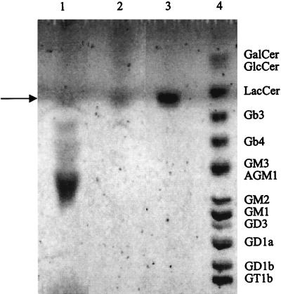
FIG. 5.
Occurrence of sulfatide-like glycolipid in pig jejunal mucus and epithelial cells. Total lipid extracts were separated by TLC developed in chloroform–methanol–0.02% CaCl2 in water (55:45:10 by volume) and visualized with Bial’s reagent. Lanes: 1, total lipids extracted from mucus of pig jejunum (200 μg); 2, total lipids extracted from epithelial cells of pig jejunum (200 μg); 3, sulfatide standard (5 μg); 4, glycolipid standards GalCer (10 μg), GlcCer (10 μg), LacCer (10 μg), Gb3 (2.5 μg), Gb4 (2.5 μg), GM3 (2.5 μg), GM2 (2.5 μg), GM1 (2.5 μg), GD3 (2.5 μg), GD1a (2.5 μg), GD1b (2.5 μg), GT1b (2.5 μg), and asialo-GM1 (2.5 μg). Reference glycolipids are indicated on the right. The arrow indicates the sulfatide. For abbreviations, see Table 1.

