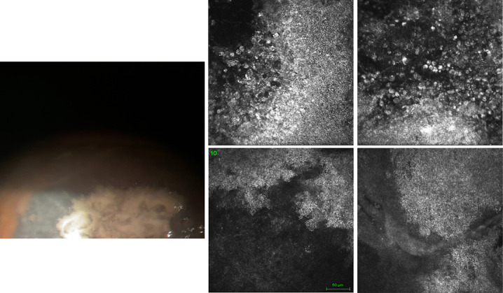
FIG. 1.

Case 1: The photograph shows the presence of rounded lesions, confluent with blurred margins, of whitish color. IVCM highlights the presence of large hyperreflecting areas formed by several spherical formations that show a caviar-like appearance, and their diameter, in some areas, gradually increases up to the edges of the lesion. IVCM, in vivo confocal microscopy.
