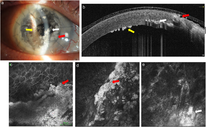
FIG. 3.
Case 2: The slitlamp and AS-OCT evaluation indicates central intrastromal whitish snowflake infiltrates (white arrow) and rounded infiltrates with jagged edges in the periphery at the level of the graft sutures (red arrow), located superficially with melted epithelium; also, endothelial deposits were present (yellow arrow) (a). The IVCM images emphasize lesions formed by spherical lesions similar to caviar, well-defined superficially (red arrow) (a–d), whereas at stromal level, the definition of white dots seems reduced (white arrow) (a–e). AS-OCT, anterior segment optical coherence tomography; IVCM, in vivo confocal microscopy.

