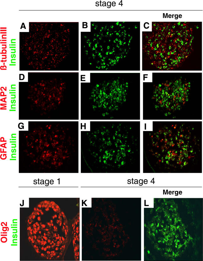
Figure 5. Stage 4 IPCs Do Not Express Markers of Differentiated Neural Cells.
(A–C) Immunohistochemical detection of β-tubulin III (A) and insulin (B) in stage 4 cells, and a merge of both images (C).
(D–F) Immunohistochemical detection of MAP2 (D) and insulin (E) in stage 4 cells, and a merge of both images (F).
(G–I) Immunohistochemical detection of GFAP (G) and insulin (H) in stage 4 cells, and a merge of both images (I).
(J–L) Immunohistochemical detction of Olig2 at stage 1 (J) and stage 4 (K) and merged view of Olig2+ and insulin+ cells at stage 4 (L). Original magnification was 630×.

