Figure 3.
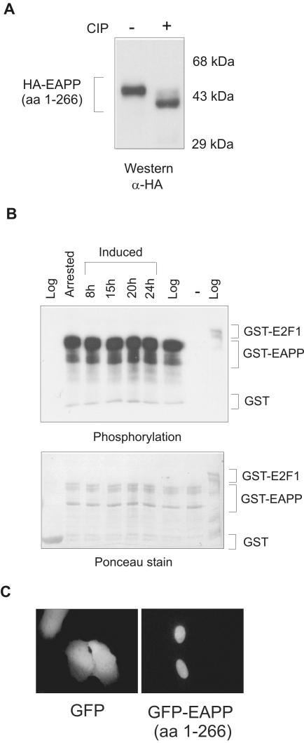
Phosphorylation and intracellular localization of EAPP. (A) HA-tagged EAPP was transiently expressed in U2OS cells. Extracts from these cells were treated with CIP (calf intestine ALP) and analyzed by SDS-PAGE, Western blotting, and immunostaining with an anti-HA antibody. (B) Glutathione-agarose bound GST, GST-EAPP, and GST-E2F-1 fusion proteins were incubated with extracts from logarithmically growing, serum-starved, and reinduced T98G cells and [γ-32P]ATP. GST served as a negative and GST-E2F-1 as a positive control for phosphorylation. Proteins were incubated for 30 min at 30°C, washed three times with GST-lysis buffer, boiled with protein loading buffer, and separated by SDS-PAGE. The gel was blotted to a nitrocellulose membrane, stained with Ponceau S, and exposed to an x-ray film. The top panel is the autoradiography and shows the phosphorylation. The bottom panel is the Ponceau stain that shows the amounts of fusion proteins. (C) GFP and GFP-EAPP (1–266) were transiently expressed in U2OS cells. Forty-eight hours after transfection, images were taken. GFP is distributed throughout the cell, whereas GFP-EAPP appears predominantly in the nucleus.

