Figure 1.
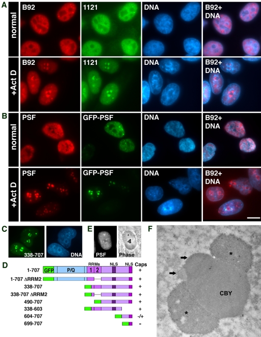
PSF and GFP-PSF localize in the same nucleolar caps. (A) Immunofluorescence images of double staining for nucleoplasmic PSF with the B92 mAb and the 1121 polyclonal antibody in untreated and in ActD-treated cells. (B) GFP-PSF–transfected cells were labeled with the B92 antibody to PSF. Hoechst DNA counterstain is shown in blue. The forth column on the right is a computer-generated overlap of the PSF and DNA stain showing the dense chromatin ring surrounding the nucleolar caps. Bar, 10 μm. (C) GFP-PSF 338–707 forms nucleolar caps. (D) Other GFP-PSF constructs were tested for nucleolar cap formation: +, form caps; -, do not form caps. P/Q, proline- and glutamine-rich domain; RRM, RNA recognition motif; NLS, nuclear localization sequences. (E) PSF (left panel: immunofluorescence) is found in phase dark nucleolar caps (DNCs; right panel: phase contrast). (F) Immunogold labeling with anti-PSF showed localization in large concave caps (stars) situated on the central body (CBY). The smaller caps (arrows) represent the segregated DFC and FC.

