Figure 6.
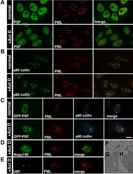
PML protein is found in distinct nucleolar caps. Confocal images of (A) Untreated: PSF was nucleoplasmic, whereas PML was found in PML bodies. Treated: PSF and PML were found in two different nucleolar caps. (B) Untreated: p80 coilin was in Cajal bodies and PML was in PML bodies. Occasionally, there was close association between the two bodies. Treated: p80 coilin and PML were in two separate caps. (C) GFP-PSF–transfected cells were labeled with antibodies to PML and p80 coilin. The three different cap structures were seen in the treated cells. Bar, 10 μm. (D) PML caps were distinct from Nopp140 LNCs and (E) UBF fibrillar caps. (F) Immunogold labeling with anti-PML detected PML bodies in the nucleoplasm of untreated cells and (G) PML in small cap structures adjacent to DNCs or (H) on top of DNCs.

