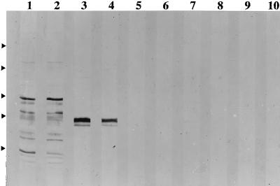
FIG. 4.
Analysis of sera from mice infected s.c. with different numbers of BCG at 6 weeks postinfection for antimycobacterial IgG1 and IgG2a antibodies. Sera were pooled from three mice similarly injected. The pattern for sera collected 6 weeks postinfection is similar to but stronger than the pattern for sera collected at 3 weeks and similar to that seen for sera collected at week 12. The odd- and even-numbered lanes were developed to reveal antimycobacterial IgG1 and IgG2a antibodies, respectively. Sera were from mice injected with 4 × 10⁸ (lanes 1 and 2), 4 × 10⁶ (lanes 3 and 4), 4 × 10⁴ (lanes 5 and 6), and 4 × 10² (lanes 7 and 8) BCG CFU and with saline (lanes 9 and 10). The relative positions of the molecular weight markers are indicated by the arrowheads at the left; from the top of the blot, the markers are maltose-binding protein–β-galactosidase (175 kDa), maltose-binding protein–paramyosin (83 kDa), glutamic dehydrogenase (62 kDa), aldolase (47.5 kDa), and triosephosphate isomerase (32.5 kDa).

