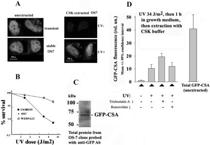
Figure 3.
Expression of GFP-CSA protein in human cells (CS3BESV) with mutation of endogenous CSA protein. (A) Images of cells expressing GFP-CSA protein. Left two images show transiently (top) or stably (bottom) transfected living cells, and right two images show unirradiated (top) or UV-irradiated (bottom) stably transfected cells preextracted with CSK buffer containing 0.5% Triton X-100 (Kamiuchi et al., 2002). Bar (A), 10 μm. UV dose was 34 J/m2 followed by 1-h incubation of cells in growth medium. (B) Survival of stably transfected GFP-CSA expressing cells (clone OS-7) after UV, parental cells (CS3BESV), and normal human fibroblasts (line WI38VA13). (C) Immunoblot of proteins from OS-7 cells probed with antibodies against GFP. (D) UV-induced insolubilization of GFP-CSA protein in OS-7 cells and its stimulation by pretreatment with trichostatin A (300 nM, 24 h). Roscovitine (20 μM) was added for 6 h before UV-irradiation. Mean GFP-CSA fluorescence intensity >50 of nuclei of OS-7 cells was measured under identical conditions (amplification gain and magnification) for all variants.

