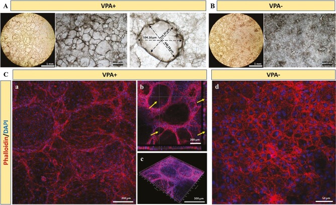
Figure 3.

The morphological characterization of the differentiated cells with or without VPA treatment. (A) Bright field images of the morphology of cholangiocyte-like cells after the treatment of VPA for 7 days. (B) Bright field images of the morphology of cells without VPA treatment. (C) Phalloidin and DAPI staining of the differentiated cells with or without VPA treatment. Arrow: biliary cystic-like structures.
