FIGURE 2:

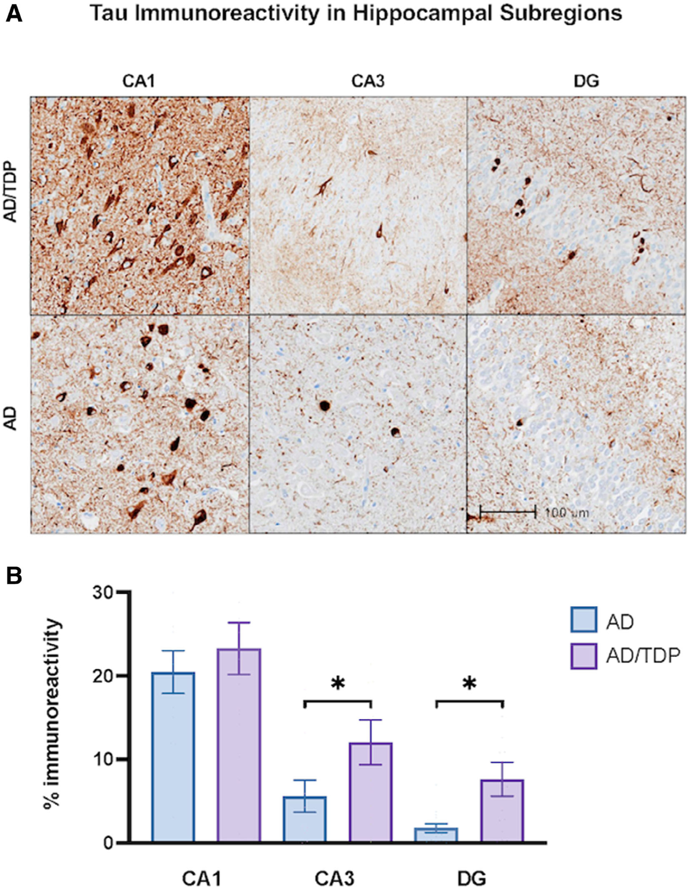
Tau immunoreactivity in amnestic dementias across subregions of the hippocampal complex. (A) Hippocampal subregions immunohistochemically stained with AT8 to visualize phosphorylated tau immunoreactivity in cases with amnestic dementia due to AD alone and due to Alzheimer’s disease (AD) with comorbid transactive response DNA-binding protein 43 (TDP-43) pathology (AD/TDP). The AD/TDP case shown is an 83-year-old woman with an 18-year history of amnestic dementia. The AD case is a 89-year-old woman with a 19-year history of amnestic dementia. Both cases were found to show distribution and extent of neurofibrillary tangle consistent with Braak stage IV/V. Images were taken at 10x magnification. (B) There was significantly more tau immunoreactivity in AD/TDP versus AD cases in both the CA3 and dentate gyrus (DG), but not the CA1. Bars represent mean percentage of area occupied by tau immunoreactivity and standard error is shown. *p < 0.05
