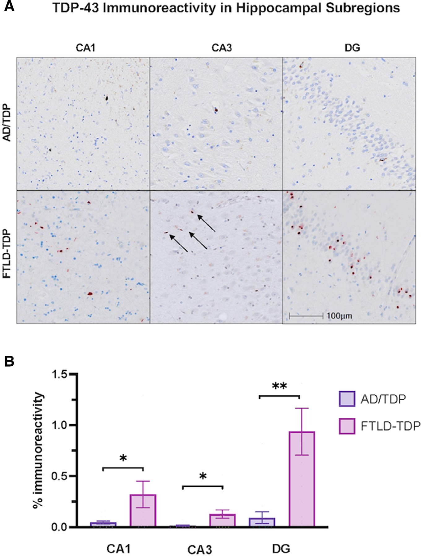
FIGURE 3:

Transactive response DNA-binding protein 43 (TDP-43) immunoreactivity across hippocampal subregions in Alzheimer’s disease (AD) with comorbid transactive response DNA-binding protein 43 (TDP-43) pathology (AD/TDP) versus TDP-43 proteinopathy associated with frontotemporal lobar degeneration (FTLD-TDP). (A) Hippocampal subregions stained with TDP-43 to visualize TDP-43 immunoreactivity in amnestic dementia due to AD/TDP and non-amnestic dementias (PPA and bvFTD) due to FTLD-TDP. The FTLD-TDP case is a 65-year-old woman with a 13-year history of primary progressive aphasia found to have FTLD-TDP type C at autopsy, and the AD/TDP case is 83-year-old woman with an 18-year history of amnestic dementia, diagnosed with Braak stage VI (see Fig 2A). Images were taken at 10x magnification. (B) There was significantly more TDP-43 positivity in FTLD-TDP across all hippocampal subregions compared with AD/TDP. Overall, TDP-43 immunoreactivity within AD/TDP was relatively low across all regions. Bars represent mean percentage of area occupied by TDP-43 immunoreactivity and standard error is shown. *p < 0.05; **p < 0.01
