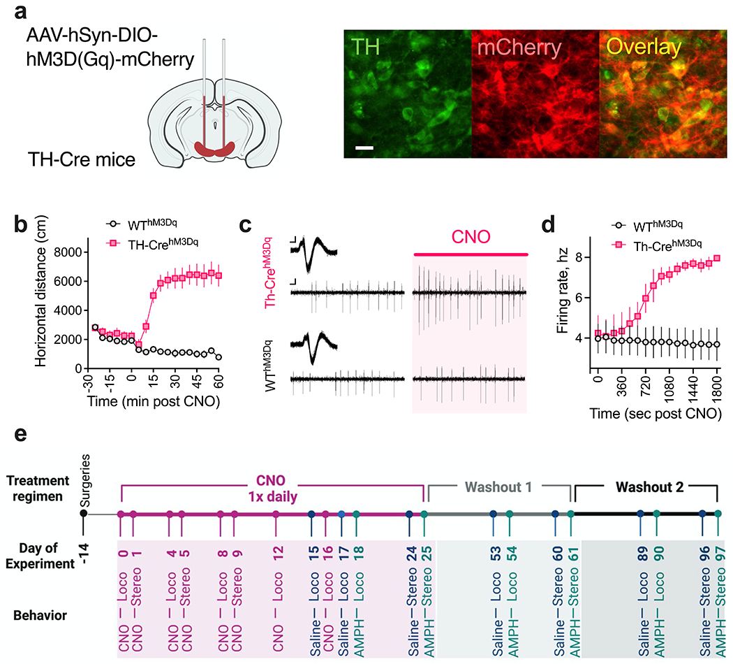
Fig. 1. Validation of DREADD-based activation of dopaminergic neurons.

(a) Schematic of AAV-hSyn-DIO-HM3D(Gq)-mCherry injections into the midbrain of TH-Cre and littermate WT control mice. Shown are representative sections from the midbrain region of TH-CrehM3Dq mice. Scale bar: 20 um. (b) Increased open-field locomotion in TH-CrehM3Dq mice following acute 1 mg/kg CNO injection. N = 8 TH-CrehM3Dq and 10 WThM3Dq mice. (c-d) Increased firing rate of DA neurons in TH-CrehM3Dq mice following acute 1 mg/kg CNO administration. (c) Representative extracellularly recorded DA neuronal waveforms (insets) and spike patterns from TH-CrehM3Dq and WThM3Dq mice before (Left) and after ~ 25 mins (Right) of CNO injection. Scale bar: Waveform, 0.05mv/0.5ms; spike patterns, 0.05mv/50ms. (d) Following a baseline recording period of 2 min, mice were administered an IP dose of 1 mg/kg CNO as recording continued for another 30 mins. N = 3 neurons/3 TH-CrehM3Dq and 3 neurons/3 WThM3Dq mice. (e) Experimental timeline for longitudinal behavior. Surgeries were conducted on P60 (Day −14). After a two-week incubation period, mice were administered an IP dose of 1 mg/kg CNO once daily P74-99. CNO was administered during the test sessions on CNO test days (for locomotion: Days 0, 4, 8, 12, 16; for stereotypy: Days 1, 5, 9) and at the conclusion of the session on saline/AMPH test days (for locomotion: Days 15, 17, 18; for stereotypy: Days 24, 25). CNO was administered to mice within their home cages at ~ the same time on non-test days. Mice were tested again for saline and AMPH responses one month (Days 53-54 and 60-61) and two months (Days 89-90 and 96-97) after stopping CNO treatment.
