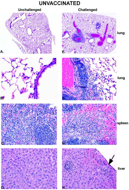
FIG. 4.
Histologic appearance of unvaccinated BALB/c mice preceding and 4 days after intranasal NMFTA1 infection. Hematoxylin- and eosin-stained tissues from unvaccinated, unchallenged (control) mice are shown on the left (A to D), and those from unvaccinated, challenged mice are shown on the right (E to H). Necrotizing inflammation is evident in all tissues from challenged mice. The arrow points to a small colony of extracellular bacteria. Approximate magnifications: ×10 (A and E) and ×200 (B to D and F to H).

