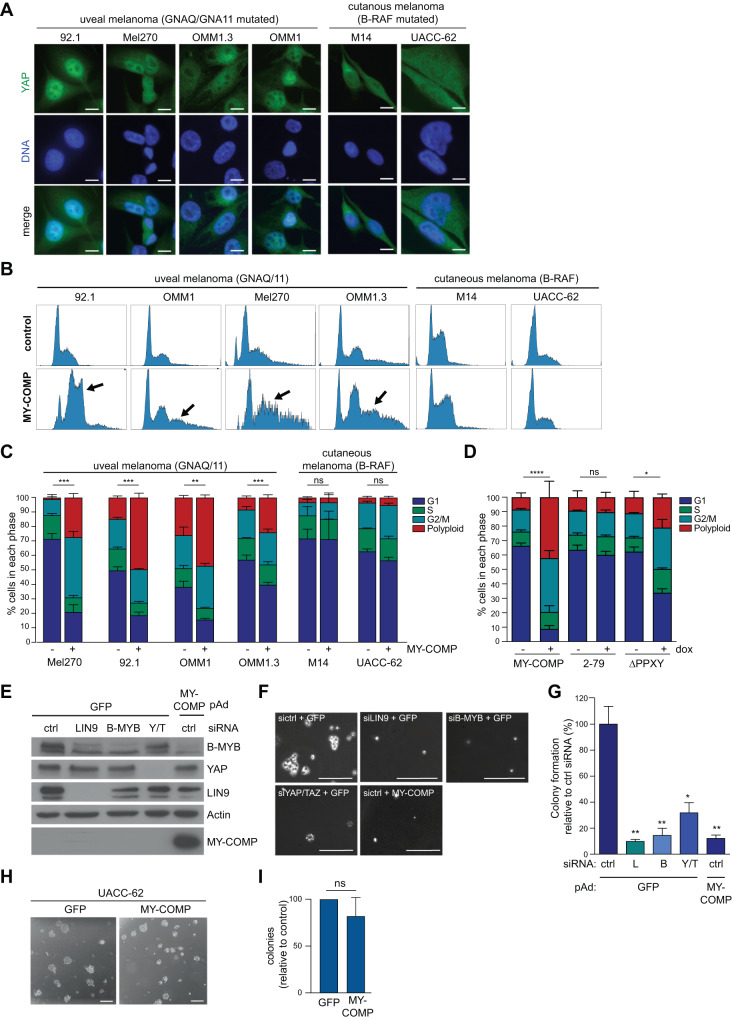
Fig. 5. Uveal melanoma cells are sensitive to inhibition of the YAP-B-MYB interaction.
A A panel of uveal melanoma cell lines (92.1, Mel271, OMM1.2 and OM1) and B-RAF mutated cutaneous melanoma cell lines (M14 and UACC-62) were immunostained for YAP. Scale bar: 10 µm. YAP localizes to the nucleus in uveal melanoma cell lines but not in B-RAF mutated cutaneous cell lines. B The cell lines described in A were stably transfected with doxycycline-inducible MY-COMP. MY-COMP was induced for 4 days and the fraction of cells in the different phases of the cell cycle was analyzed by FACS. C Quantification of the FACS data obtained from three independent experiments. Error bars depict SEM. Student’s t test was used to calculate the significance of the differences in polyploid cells. **p < 0.01, ***p < 0.001, ns not significant. D 92.1 cells were stably transfected with the indicated inducible MY-COMP constructs coupled to GFP through a T2A site. FACS was performed to determine the percentage of in different phases of the cell cycle without or with induction of the constructs. After induction, GFP-positive cells were analyzed. Error bars indicate SDs. n = 3 independent replicates. One way ANOVA. E 92.1 cells were transfected with the indicated siRNAs directed at LIN9, B-MYB or YAP/TAZ or were infected with a recombinant adenovirus encoding MY-COMP. As control, cells were transfected with a siRNA against luciferase and infected with an adenovirus encoding GFP. Expression of the indicated proteins was investigated by immunoblotting. Actin served as a control. F, G To assay for anchorage-independent growth, cells as described in E were seeded in 0.6% agarose and grown for 21 days. F shows representative images (scale bar: 25 µm) and G shows a quantification of one experiment performed in triplicate. siRNAs: L = LIN9, B = B-MYB, Y/T = YAP and TAZ. Error bars indicate SEM. H, I To assay for anchorage-independent growth, UACC-62 cells were infected with a recombinant adenovirus encoding MY-COMP or with GFP as a control. Infected cells were then seeded in 0.6% agarose and grown for 14 days. H shows representative images (scale bar: 25 µm) and I shows a quantification of one experiment performed in triplicate. Error bars indicate SD. n = 2.

