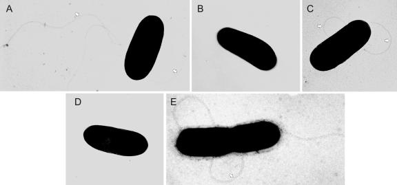
FIG. 3.
Electron micrographs of the flagellated L. monocytogenes wild-type (WT) strain EGD (A and C) and the nonflagellated ΔdegU mutant (B and D). The flagellated but nonmotile ΔcheY mutant is included as a control (E). Bacteria were grown at 24°C (A, B, and E) or 37°C (C and D) and examined under a transmission electron microscope (TEM 100; Zeiss) after negative staining with 0.5% uranyl acetate for 1 min. The flagella present in the WT and ΔcheY strain are clearly visible (arrowheads). Original magnification: 20,000-fold.

