
Fig. 4. Midgut progenitors actively eliminates Plasmodium oocysts and internalize parasite remnants.
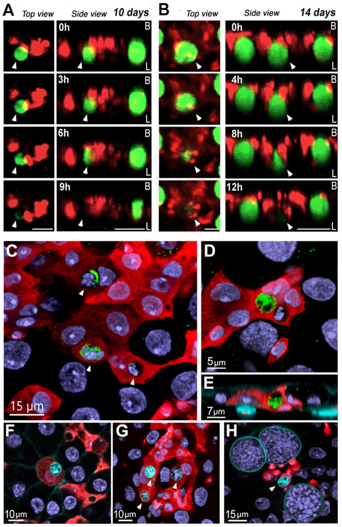
A Whole mosquito live imaging over the course of 9 h 10 days pi. Scale bar from top view: 20 µm and from side view: 30 µm. B Whole mosquito live imaging over the course of 12 h 14 days PI. Scale bar from top view: 20 µm and from side view: 50 µm. C Oocyst fragments inside midgut progenitors at 5 days PI. White arrows indicate parasite remnants inside of midgut progenitors. Scale Bar: 15 µm. Leftover of P.berghei GFP from inside of a midgut progenitor 5 days PI (D) Front view and (E) Lateral view. Scale Bar: 5 µm and 7 µm respectively. Midgut progenitors are in red and P.berghei are in green (F) Dead oocyst in cyan inside of a midgut progenitor 5 days PI. Scale Bar: 10 µm. G Dead oocysts in cyan inside midgut progenitors 5 days PI. Scale Bar: 10 µm. H Live oocysts with cyan staining on the surface and a smaller dead oocyst with staining inside the capsule, engulfed by a midgut progenitor. White arrows indicate dead oocysts. Scale Bar: 15 µm. Micrographs are representative of experiments that were independently reproduced at least 2 times with multiple individuals in each experimental group and obtained similar results.