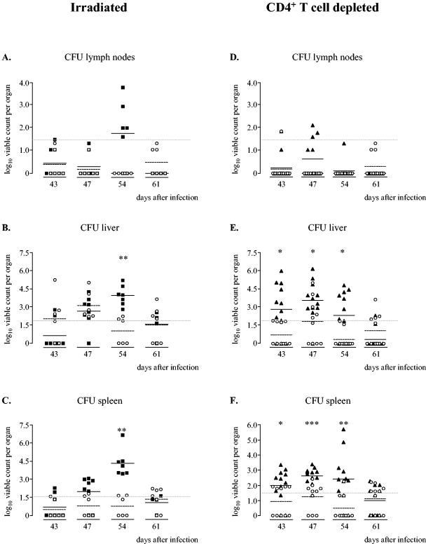
FIG. 1.
Bacterial loads within the lymph nodes (A), livers (B), and spleens (C) of mice infected with S. enterica serovar Typhimurium 14028s that were irradiated on day 41 (▪) and untreated infection controls (○) and bacterial loads within the lymph nodes (D), livers (E), and spleens (F) of mice depleted of CD4+ T cells (▴) and infection controls (○). On days 39, 41, and 43 after infection, the mice were injected i.p. with 200 μg, 100 μg, and 100 μg of rat anti-CD4 GK1.5 antibody, respectively. The infection controls were injected i.p. with an equal volume of PBS. At different times, livers, spleens, and lymph nodes were aseptically removed, and cell lysates were made. The viable counts in the organs were determined by plating serial dilutions of the cell lysates and are expressed as log10 viable counts (means ± standard errors of the means). Data from two independently performed experiments are shown. Asterisks indicate statistically significant differences compared to the infection controls (one asterisk, P < 0.05; two asterisks, P < 0.005; Mann-Whitney rank order test), and the gray dashed lines indicate the detection limits of the microbiological method (50 CFU for the livers and 30 CFU for the spleens and lymph nodes).

