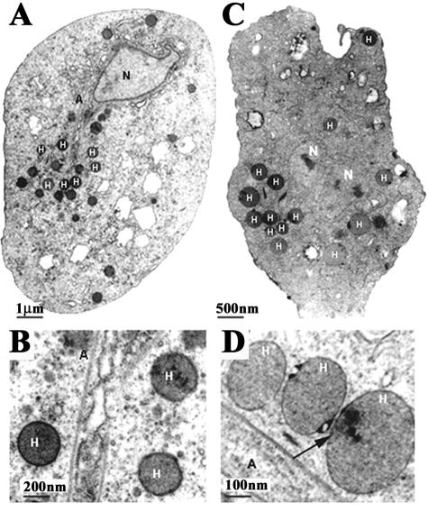
FIG. 2.
Transmission electron microscopy showing comparative thin sections and ultrastructure of control (A and B) versus 20 mM DAB-treated (C and D) T. vaginalis isolate T016. The hydrogenosomes (H), axostyle (A), and nucleus (N) are readily visible as indicated. All of the structures present a normal appearance. A higher magnification of T. vaginalis to visualize hydrogenosomes in routine preparations is shown in panels B and D. Notice the normal appearance of all cell structures, including the hydrogenosomes (H), in the DAB-treated organisms (C and D), although, on occasion, there were electron-dense spots in the hydrogenosomal matrix (panel D, arrow).

