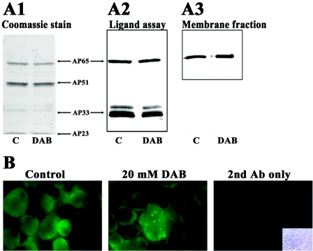
FIG. 3.
Trichomonads treated with 20 mM DAB express identical amounts of adhesins as control, untreated parasites. (A) Panel A2 shows immunoblots of a HeLa cell-binding proteins from a ligand assay (Materials and Methods) with AP65 and AP33 detected with MAbs 12G4 and F5.2, respectively (21, 24). The left panel (A1) has duplicate Coomassie brilliant blue-stained gels after the ligand assay as in the right panel and shows the other two AP51 and AP23 adhesins (14, 34). Multiple bands for AP33 in panel A2 are due to partial degradation by the trichomonad cysteine proteinases, as described previously (21). Panel A3 shows immunoblot results for AP65 as in A2 of a ligand assay performed by using enriched membranes from identical cell equivalents of control, untreated and 20 mM DAB-treated parasites. (B) Immunofluorescence was performed with nonpermeabilized trichomonads with MAb 12G4, which reacts with surface AP65, as demonstrated recently (24, 38). No fluorescence was obtained in the absence of anti-AP65 MAb but in the presence of fluorescein-conjugated secondary goat anti-mouse IgG, as shown in the far right panel. The inset (bottom right) in the far right panel shows the bright-field microscopy picture of the cells used in the secondary antibody control.

