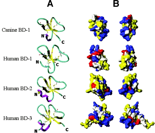
FIG. 3.
Secondary structure assignment and surface analysis for human and canine β-defensins. (A) The leftmost column depicts ribbon diagrams showing α-helix (purple), β-sheet (yellow), and coil (turquoise) secondary structure elements for the four peptides. (B) The two rightmost columns depict front and back views of peptide solvent accessible surfaces. Acidic (anionic) residues are red, basic (cationic) residues are blue, and hydrophobic residues are yellow.

