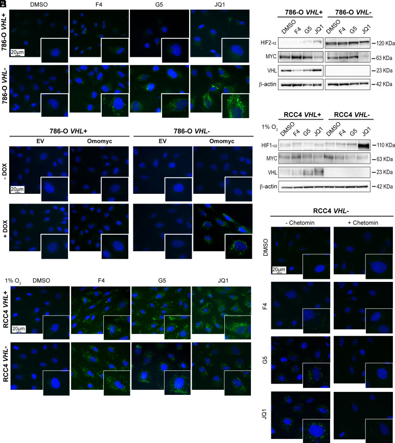
Fig. 1.
HIF stabilization is required for LD accumulation upon MYC inhibition in ccRCC cells. (A) Lipid droplet staining and (B) western blot of the indicated proteins in 786-O VHL+ and VHL− cells after MYC inhibition. (C) Lipid droplet staining in 786-O VHL+ EV, 786-O VHL+ Omomyc, 786-O VHL- EV, or 786-O VHL- Omomyc cells after vehicle (−Omomyc, H2O) or DOX treatment (+Omomyc). (D) Western blot of the indicated proteins in RCC4 VHL+ and VHL− cells after treatment with MYCis in 1% O2 (hypoxia). (E) Lipid droplet staining in RCC4 VHL+ and VHL− cells after treatment with MYCis in 1% O2 (hypoxia). (F) Lipid droplet staining in RCC4 VHL+ and RCC4 VHL− cells after treatment with MYCis in combination with DMSO (−chetomin) or chetomin. For western blots, β-actin was used as loading control. Molecular weight markers are shown to the Right. For fluorescence images: blue, DAPI; green, LD-BTD1 (LDs). (Scale bars: 20 μm.) All results are representative of three independent replicates.

