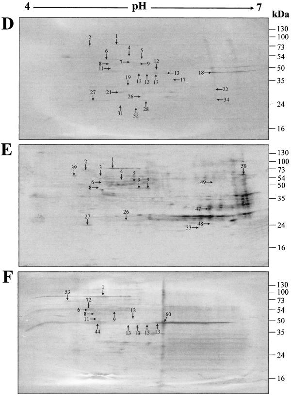
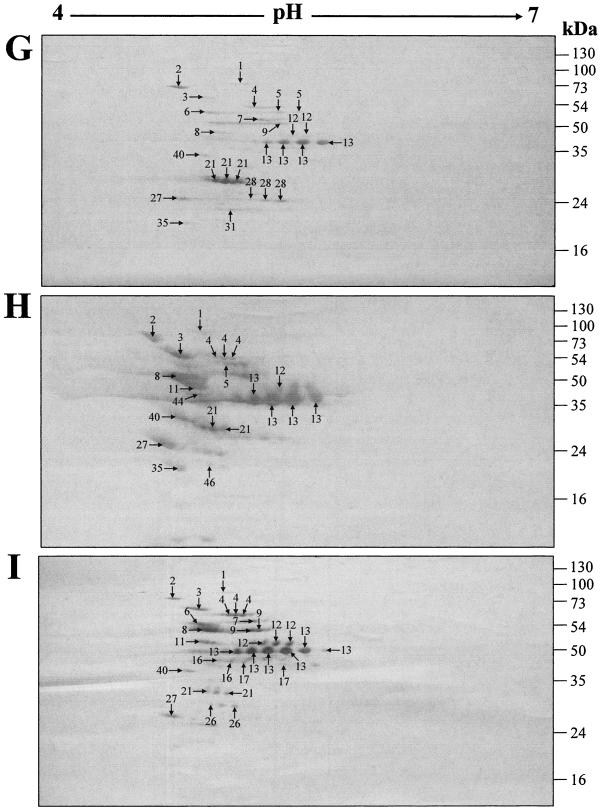
FIG. 1.
Two-dimensional gel electrophoresis profiles of GAS mutanolysin cell wall extracts. The extracts were harvested from GAS strains NS931 (A, D, and G), NS13 (B, E, and H), and S43 (C, F, and I) after growth to late stationary phase (37°C for 16 h) in Todd-Hewitt medium (Difco) supplemented with 1% (wt/vol) yeast extract without shaking. The protein extracts (170 μg) were isoelectric focused over a linear pH gradient of 4 to 7 and resolved with a 12.5% SDS-PAGE gel. (A-C) The gels were stained with colloidal Coomassie blue and destained in 1%anti-human IgG-HRP conjugate (Bio-Rad). Negative-control blots probed only with goat anti-human IgG-HRP conjugate contained no immunoreactive proteins (result not shown). (G-I) The cell surface of each strain was labeled with biotin before the mutanolysin extract was harvested. The proteins were transferred to a PVDF membrane and probed with an SA-HRP conjugate prior to development with diaminobenzidine. Negative-control blots of nonbiotinylated extracts contained no labeled proteins (result not shown). Protein spots identified by peptide mass (vol/vol) acetic acid. (D-F) The proteins were transferred to a PVDF membrane and probed with a 1:100 dilution of pooled human sera from an area of endemicity. Bound antibodies were detected using a goat fingerprinting are denoted by numbered arrows, which correspond to the proteins in Table 1. Molecular mass markers are given in kilodaltons.


