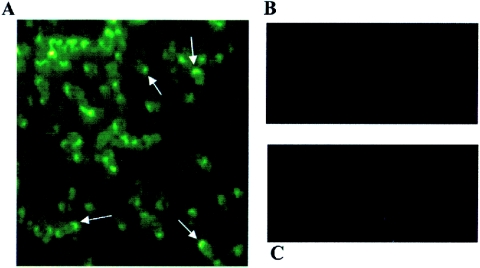
FIG. 7.
Immunofluorescent staining of Bartonella cells. (A) B. vinsonii subsp. arupensis stained with anti-Bva 6.1; (B) B. quintana stained with anti-Bva 6.1; (C) B. vinsonii subsp. arupensis stained with preimmunized mouse serum. Arrows in panel A point to the concentrated staining in the polar regions of the cells. Magnification, ×1,000.

