FIG. 3.
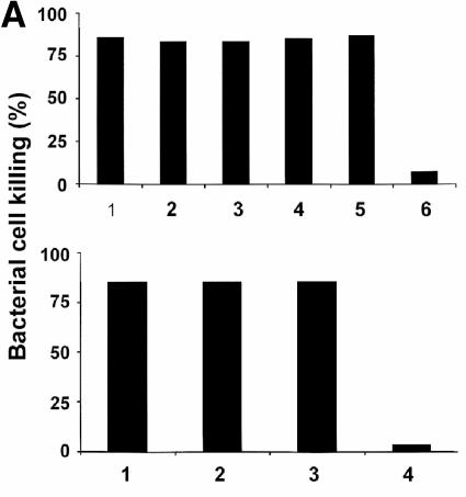
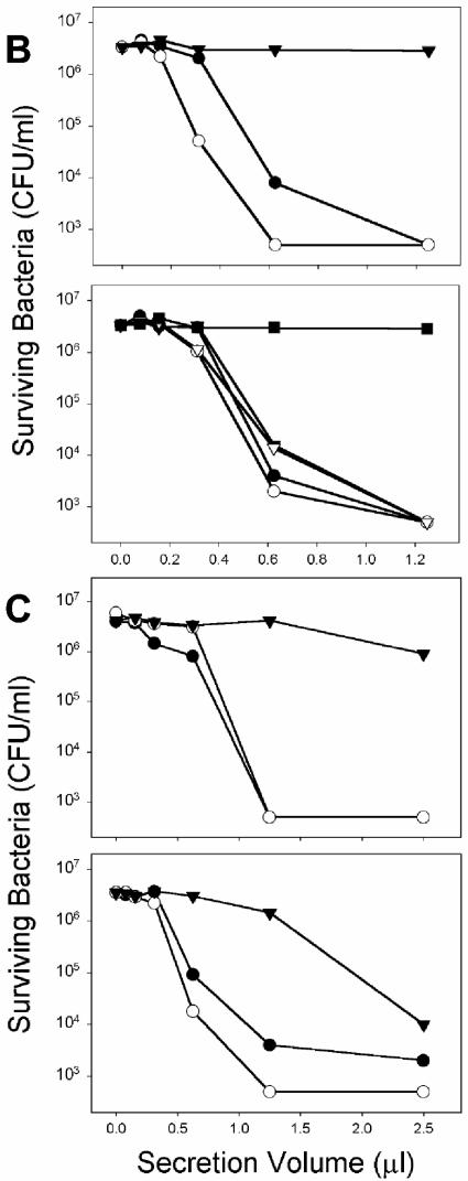
Microbicidal activity secreted from Paneth cells in response to diverse LPS, lipid A, and LTA molecules. (A) Ten-microliter samples collected from crypts exposed to WT and ΔmsbB strains of E. coli JM83 and H16 were combined with 1,000 CFU of ΔphoP serovar Typhimurium cells resuspended in 40 μl of PIPES buffer. After 60 min at 37°C, surviving CFU were quantitated by growth on semisolid media at 37°C overnight as described in the legend to Fig. 1. Groups were normalized by expressing bacterial cell killing as a percentage relative to bacteria incubated for 1 h at 37°C in iPIPES alone. Shown in the upper panel are results for E. coli LPS from Sigma (bar 1), WT JM83 (bar 2), ΔmsbB JM83 (bar 3), WT H16 (bar 4), and ΔmsbB H16 (bar 5). Also shown are results for control crypts incubated in iPIPES without antigen (bar 6). In the lower panel, results are shown for LPS from P. gingivalis (bar 1), B. forsythus (bar 2), and E. coli (Sigma) (bar 3). Also shown are results for control crypts incubated in iPIPES without antigen (bar 4). (B and C) Serial dilutions of supernatants collected from stimulated crypts exposed to purified antigens were assayed for bactericidal activity against ΔphoP serovar Typhimurium cells (see Materials and Methods). The results are representative of duplicate assays performed on separate days. (B) In the upper panel, results for secretions from crypts exposed to Mg2+-precipitated serovar Typhimurium LPS from phoP(Con) (•) and ΔphoP (○) cells and from control crypts incubated in iPIPES without antigen (▾) are shown. In the lower panel, results for secretions from crypts exposed to phenol-extracted serovar Typhimurium LPS from WT (•), WT ΔwaaP (○), phoP(Con) (▾), and phoP(Con) ΔwaaP (▿) cells and from control crypts incubated in iPIPES without antigen (▪) are shown. (C) The upper panel shows results from secretions from crypts exposed to P. aeruginosa lipid A from PAK cells grown in a low concentration of Mg2+ (•) or a high concentration of Mg2+ (○) and from control crypts incubated in iPIPES without antigen (▾). The lower panel shows results from secretions from crypts exposed to L. monocytogenes LTA from WT strain 10403S grown in a low (•) or high (○) concentration of Mg2+ and from control crypts incubated in iPIPES without antigen (▾).


