FIG. 6.
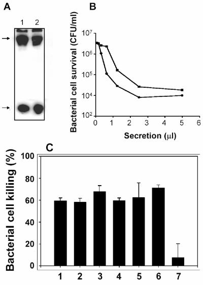
Tlr4-null Paneth cells from C3H/HeJ mice release bactericidal peptide activity in response to bacterial antigens. (A) Intestinal proteins (≤10 kDa) prepared from outbred Swiss (lane 1) and C3H/HeJ (lane 2) mice by Centricon 30 centrifugal filtration were separated by acid-urea polyacrylamide gel electrophoresis, transferred to a nitrocellulose membrane, and probed with anti-cryptdin-1 rabbit serum (2, 3). Arrows at the left denote the presence of immunoreactive procryptdin (upper arrow) and mature cryptdins 1 through 3 and 6 (lower arrow), respectively. (B) Approximately 5,000 crypts were incubated with 103 CFU of ΔphoP serovar Typhimurium cells/crypt for 30 min at 37°C in 500 μl of iPIPES. Samples of collected secretions were assayed for bactericidal activity against 5 × 10⁶ CFU of ΔphoP serovar Typhimurium cells. The release of abundant bactericidal peptide activity by crypts from C3H/HeJ mice is evidence that Paneth cell secretory responses to bacteria are not mediated by Tlr4. (C) Solid bars denote the bactericidal activity of secretions released by C3H/HeJ mouse small intestinal crypts exposed to LPS (100 ng/ml) from P. aeruginosa PAK cells grown in 8 μM MgCl2 (bar 1), LPS (100 ng/ml) from P. aeruginosa ΔphoP PAK cells grown in 8 μM MgCl2 (bar 2), LPS (100 ng/ml) from ΔphoP serovar Typhimurium (bar 3), lipid A (1 μg/ml) from ΔphoP serovar Typhimurium (bar 4), LTA (10 μg/ml) from WT L. monocytogenes Scott A-M (see Materials and Methods) (bar 5), commercially available LPS (Sigma, 100 ng/ml) from serovar Typhimurium (bar 6), and control crypts incubated in iPIPES without antigen (bar 7).

