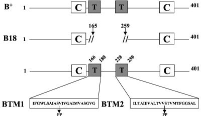
FIG. 1.
Schematic diagram of YopB primary structure and mutant design. The structure of wild-type YopB protein (B+) is indicated at the top. Boxes represent structural elements, corresponding to predicted transmembrane domains (T) and coiled-coil (C) regions. The middle structure represents YopB18 (B18), which results from an in-frame deletion of amino acids 166 to 258, removing both transmembrane domains. The lower structure shows the transmembrane domains expanded to single-letter code, and the positions of double proline substitutions are indicated. YopBTM1 (BTM1) contains prolines substituted for serine 175 and valine 176. YopBTM2 (BTM2) contains prolines substituted for valine 239 and serine 240.

