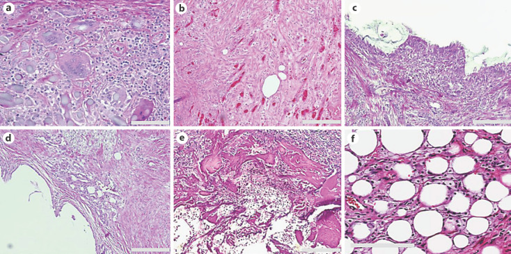
Fig. 1.
H&E images of common histologic changes observed around the needle tract. a Giant-cell reaction with macrophage aggregation. b Granulation tissue with the presence of fibroblasts. c Metaplastic synovial mucosa lining the biopsy cavity. d Vascular-rich granulation tissue. e Residual carrier material from biopsy marker inside the biopsy cavity. f Fat necrosis. Scale bars, 200 μm.

