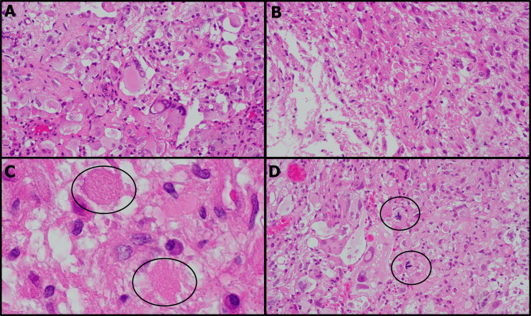
Figure 5. H&E stained histology images of the excised specimen.
The histological specimen displayed characteristics of pleomorphic xanthoastrocytoma. Most nuclei were enlarged and exhibited bizarre shapes, accompanied by a vacuous, foamy cytoplasm (A). A significant number of xanthic cells were observed (B); numerous eosinophilic granular bodies were also visible (C). Mitotic figures are present (D).

