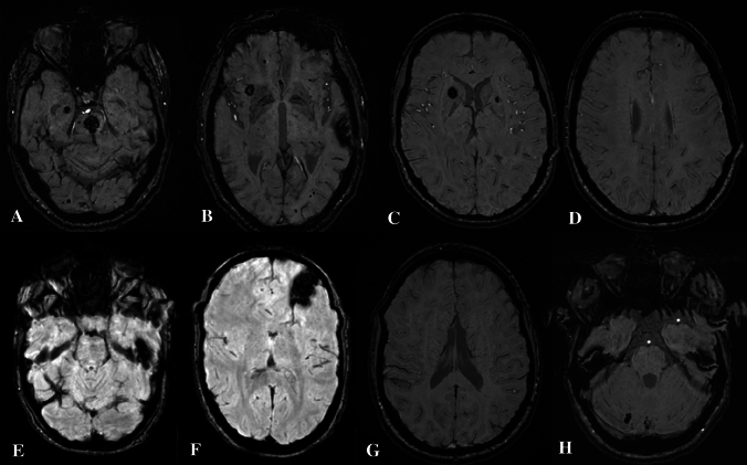
Fig. 3.
Susceptibility-weighted imaging, axial planes of the four members of the family. A, B Proband (II-2), multiple cavernomas, supra and infratentorial, the largest ones in pons (A) and left superior temporal lobe (B) and numerous type IV cavernomas. C, D Older daughter (III-3), right caudate nucleus/anterior limb of internal capsule and left lenticular nucleus (C), infracentimetric cavernomas located in right supramarginal gyrus (D), and additional type IV cavernomas mainly in the frontal region. E, F Younger daughter (III-5), two major cavernomas on left temporal superior gyrus (E) and fronto-orbitary region (F) and a pericentrimetric cavernoma with acute hemorrhage and size increasing in the left superior lobe parietal lobe. G, H Granddaughter (IV-2), one cavernoma on right cerebellum, without acute hemorrhage. No visible type IV cavernomas

