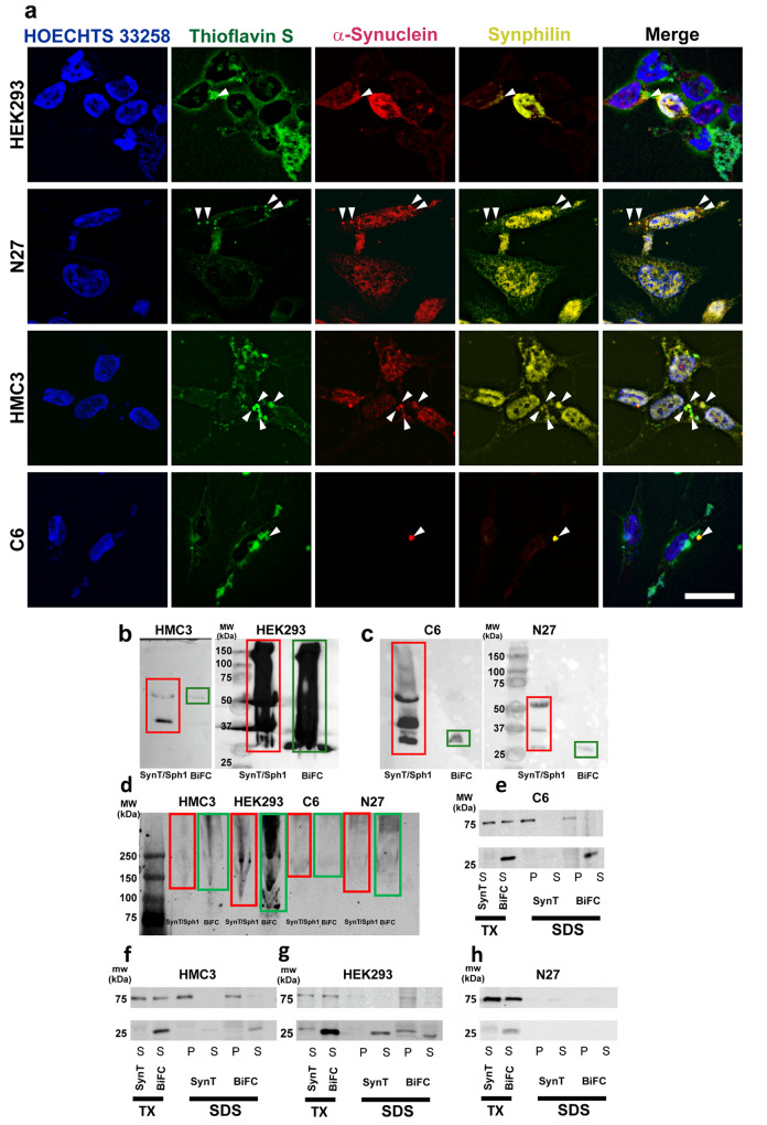
Fig. 2. Formation of inclusions in our in vitro methods.
a Representative pictures of HEK293, N27, HMC3, and C6 cells showing thioflavin S-positive staining for aggregates (green) α-synuclein-T (red), V5-synphilin-1 (yellow) immunoreactivity. Arrows point at possible Lewy body-like structures. Scale bar 15 µm. b SDD-Tricine membrane of HMC3 (left side) and HEK293 (right side) cells transfected with α-synuclein-T and V5-synphilin-1 (SynT/Sph1, red rectangle), VN-α-synuclein and α-synuclein-VC (BiFC, green rectangle). HMC3 cells show two bands of ~40 and 60 KDa when transfected with α-synuclein-T, which may be composed of α-synuclein dimers and trimers. HEK293 cells show a smear of different MW ranging from ~25 KDa to more than 150 KDa. c SDD-Tricine membrane of C6 (left side) and N27 (right side) cell lines transfected with SynT/Sph1 (red rectangle), VN-α-synuclein and α-synuclein-VC BiFC (green rectangle). C6 cells show several bands that may be oligomers. N27 cells show the presence of possible monomers, dimers, and trimers. d Native WB membrane of C6, N27, HMC3, and HEK293 cell lines transfected with SynT/Sph1 (red rectangle), VN-α-synuclein and α-synuclein-VC BiFC (green rectangle). e–g SDS-PAGE WB membranes for Triton-X and SDS solubility assays in C6, HMC3, and HEK293 cells. BiFC and SynT/Sph1 models show Triton-X soluble signals at high molecular weight. h SDS-PAGE WB membranes for Triton-X and SDS solubility assays in N27 cells. No signal has been observed in SDS-soluble fractions after Triton-X digestion (N = 4 independent experiments). TX: Triton-X. P: Pellet (insoluble) fraction. S: Soluble fraction.

