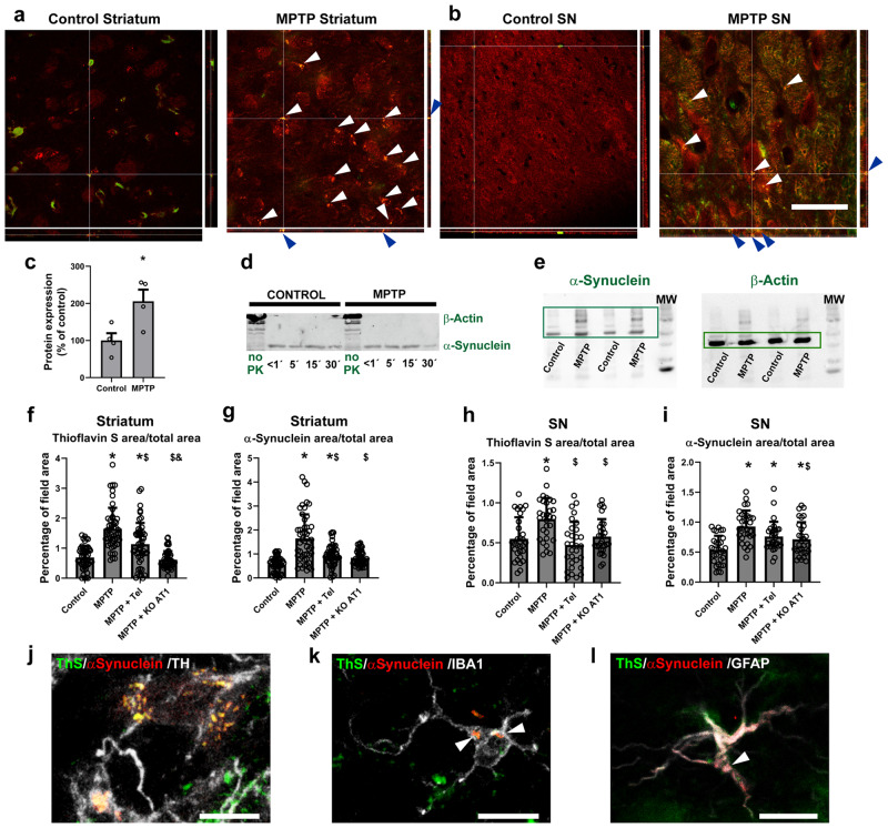
Fig. 6. Striatum and substantia nigra of control mice and MPTP-treated mice.
a, b Treatment with MPTP induced the formation of inclusions in the striatum (a) and nigra (b), as observed in confocal pictures with projections in all three dimensions, where white and blue arrows point to the inclusions. Scale bar: 50 µm; Red: α-synuclein. Green: ThS. c Treatment with MPTP led to higher levels of α-synuclein oligomer phosphorylation at the Serine 129 as measured by western blot. d Treatment with MPTP did not change the resiliency of aggregated α-synuclein oligomers to pK digestion. e Treatment with MPTP induced the formation of higher molecular weight species in mouse substantia nigra as observed by SDD-tricine western blot. f, i Treatment with MPTP led to significant increase in thioflavin S-positive staining (f, h) and in α-synuclein expression (g, i) in the striatum (f, g) and substantia nigra (h, i), which were inhibited by treatment with the AT1 blocker telmisartan or the absence of AT1 receptors (AT1 KO mice). N = 50 (f, h) or 30 (i) pictures analyzed. j–l Double immunofluorescence and ThS staining of mouse tissue showing the presence of α-synuclein aggregates in nigral dopaminergic neurons (j), microglia (k), and astrocytes (l). Scale bar: 10 µm. j–l Tel: Telmisartan. MPTP + KO AT1: MPTP-treated AT1 KO mice. Red: α-synuclein. White: TH (j), IBA-1 (k), GFAP (l). Green: ThS aggregates. *p < 0.05 relative to control. $p < 0.05 relative to MPTP. &p < 0.05 relative to MPTP + Telmisartan. Unpaired two-tailed t test (c). Two one-way ANOVA and Tukey’s multiple comparison test (j–l). Error bars represent SEM.

