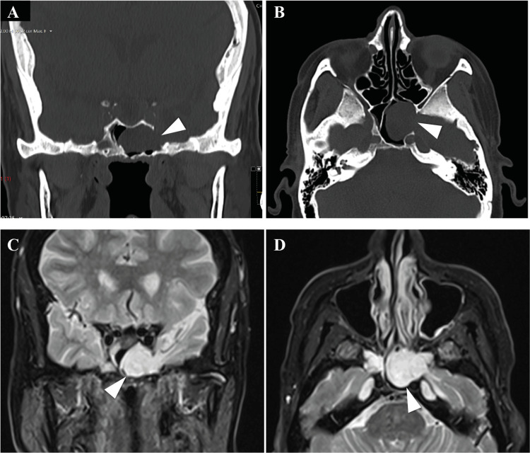
Figure 4. CT and MRI show dehiscence of the lateral left sphenoid wall with brain herniation.
CT scans (A and B) show dehiscence of the lateral left sphenoid wall (arrowheads), establishing communication with the middle cranial fossa. MRI (C and D) shows bone discontinuity in the left lateral wall of the sphenoid, crossed by tissue with signal emission characteristics similar to brain parenchyma and CSF to the left chamber of the sphenoid sinus (arrowheads).

