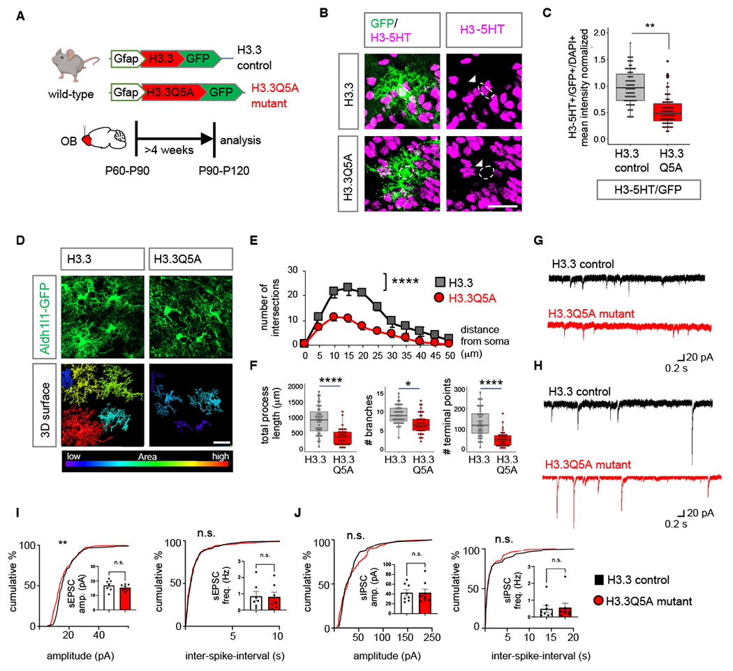
Fig. 7. Inhibition of H3-5HT in OB astrocytes disrupts astrocyte morphology.

(A) Schematic illustrating viral vectors used for H3.3 and H3.3Q5A expression in OB astrocytes. (B) H3-5HT and GFP co-labeling in H3.3 and H3.3Q5A expressing OBs and (C) box plots depicting quantification of astrocytic H3-5HT (99-110 cells/cohort, **p= 0.0092; unpaired Student’s two-tailed t-test on n= 4 mice/cohort). Scale bar: 25 µm. (D) High-magnification confocal images of H3.3-GFP and 3D surface rendering of the same showing reduced astrocyte morphological complexity in H3.3Q5A OB astrocytes. Scale bar: 20 µm. (E) Sholl analysis of astrocyte complexity (n= 4, average of 44 cells/cohort, ****p= 1.9e-05; two-way repeated measures ANOVA with Sidak correction). Data presented as mean ± SEM. (F) Quantification of total process length, branch number, and terminal points (44 cells/cohort, ****p= 2.75e-05, *p= 0.0122, ****p= 8.73e-06, unpaired Student’s two-tailed t-test on n= 4 mice/cohort). (G-H) Traces and (I-J) summary data of amplitude and frequency from (I) sEPSC recordings (7 cells/cohort, sEPSC amplitude p= 0.2386; sEPSC frequency p= 0.7917; unpaired two-tailed Student’s t-test on n= 3 mice/cohort, **p= 0.0059 K-S test); and from (J) sIPSC recordings (8 cells/cohort, sIPSC amplitude p= 0.8277, sIPSC frequency p= 0.7128, unpaired two-tailed Student’s t-test on n= 3 mice/cohort. All recordings are in granule cells from H3.3 and H3.3Q5A OB’s and data is presented as mean ± SEM. See Table S3 for data summary.
