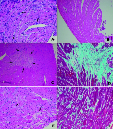
FIG. 2.
Representative sections of myocardium of mice infected with T. cruzi. (A) Day 15 p.i. in cardiac myocyte-specific ET-1 KO mice infected with the Tulahuen strain. There is myonecrosis, inflammation, vasculitis, and parasite pseudocysts. Mice infected with the Brazil strain showed similar pathology. (B) Mice infected with the Brazil strain at 130 days p.i. There is dilation of the left ventricle in infected FLOX mice. (C and D) Cardiac myocyte hypertrophy and areas of replacement (arrows) and interstitial fibrosis in Brazil strain infected FLOX mice day 130 p.i., which is more evident by Trichrome staining, as seen in panel D. (E and F) Day 130 p.i. Brazil strain-infected ET-1flox/flox;α-MHC-Cre(+) mice. Note the interstitial fibrosis, which is more evident by Trichrome staining in panel F. The genotype of the cardiac myocyte-specific ET-1 KO mice is ET-1flox/flox;α-MHC-Cre(+) and of the FLOX mice is ET-1flox/flox;Cre(−).

