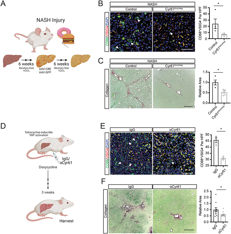
Figure 7. Blocking Cyr61 in YAP-induced Fibrosis Blunts Macrophage Recruitment and Fibrosis.
(A) Cartoon of Cyr616wkΔHep intervention after 6 weeks of NASH diet. (B) Representative immunofluorescence imaging of specified proteins in control (n=6) or Cyr616wkΔHep-intervened (n=6) NASH livers. Scale bar=100μm. Quantification of CD68+VSIG4− macrophages to the right. HPF=high power field. Arrows indicate CD68+VSIG4− cells. (D) Cartoon of tetracycline-inducible YAP liver fibrosis (YAP-Tg) with αCyr61 treatment. (D) Representative immunofluorescence imaging of specified proteins in YAP-Tg livers treated with IgG (n=3) or αCyr61 (n=3). Scale bar=100μm. Quantification of CD68+VSIG4− macrophages to the right. HPF=high power field. Arrows indicate CD68+VSIG4− cells. (F) Representative picrosirius red staining of livers from YAP-Tg mice treated with IgG (n=11) or αCyr61 (n=13) antibody. Scale bar=500μm. Quantification to the right. Mean and SEM plotted; p values calculated with Mann-Whitney U test. *p<0.05, **p<0.01, ***p<0.001.

