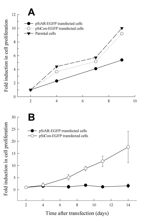
Figure 3.
Suppression of LNCaP cell proliferation in the absence of endogenous AR. LNCaP cells were seeded in tissue culture plates and transfected with a mixture of either pSiAR-EGFP or pSiCon-EGFP plasmid construct with Lipofecamine 2000 in OPTI-MEM. (A) Cell proliferation without separation of GFP-positive and negative cells. At 24 hours following transfection, cells were trypsinized and distributed into each well (1,000 cells/well) of 96-well tissue culture plates in the presence the complete medium. Cell proliferation was determined using the XTT assay kit for a period of 9 days; data from days 11 and 14 were not included since parental and pSiCon-EGFP transfected LNCaP cells reached confluence after day9. (B) LNCaP cell proliferation following enrichment of GFP-positive cells. At 24 hours after transfection, cells were trypsinized and EGFP-positive cells were collected through the MoStar cell sorting system. GFP-positive cells were seeded into each well (1,000 cells/well) of 96-well plates for cell viability assay. Cell proliferation was determined for a period of 14 days. Data were calculated as absorbance at days following transfection normalized to the absorbance at the day of cell sorting, and presented as fold induction in absorbance. LNCaP cells transfected with pSiCon-EGFP plasmid construct were used as the AR-positive control. * represents significant statistical difference between LNCaP cells with and without AR (P < 0.001). Each time point represents the mean ± SEM from 3 independent experiments.

