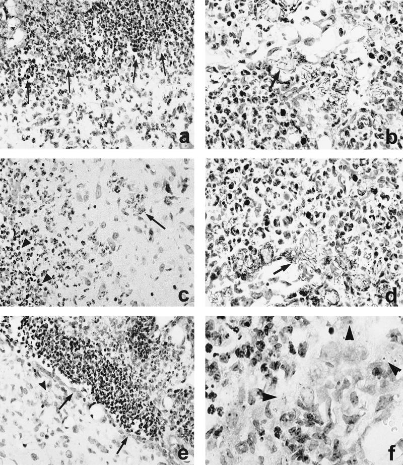
FIG. 3.
CNS pathology of nonimmunized mice i.c. infected with L. monocytogenes WT (a and b), ΔinlAB2 (c and d), and ΔplcB2 (e and f) at day 3 p.i. (a) Severe empyema of the ventricle (area above the arrows). The ependymal wall is largely destroyed and barely discernible, and the periventricular brain stem is infiltrated by numerous neutrophils and macrophages. (b) L. monocytogenes WT cluster in the largely destroyed plexus. The arrow points to a group of remarkably elongated WT bacteria. (c) ΔinlAB2 has also largely destroyed the ependyma of the ventricular wall (area left of the arrowheads) and has invaded the adjacent brain parenchyma. Brain stem neurons are surrounded by inflammatory leukocytes (arrow). (d) ΔinlAB2 cluster in the partially necrotic choroid plexus. Note that the elongated shape of the ΔinlAB2 strain (arrow) is identical to that of the WT in panel b. (e) ΔplcB2 has also infected the fourth ventricle, and the bacteria are accompanied by intraventricular neutrophils and macrophages. In contrast to WT (a) and ΔinlAB2 (c), the ependymal lining is largely intact (arrows). Very few bacteria are detectable in the periventricular tissue (arrowhead). (f) Some ΔplcB2 are detectable either as single or small groups of coccoid bacteria (arrowheads) in the choroid plexus. The choroid plexus is infiltrated by neutrophils and macrophages, but in contrast to the images shown in panels b and d, its structure is largely preserved. Specimens in panels a to f were stained with cresyl violet. Magnification is as follows: for panels a and e, ×260; b and d, ×1,250; c, ×520; f, ×1,470.

