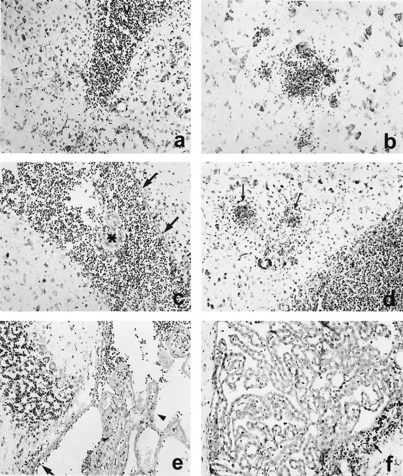
FIG. 4.
CNS pathology of immunized mice i.c. infected with L. monocytogenes WT (a and b), ΔinlAB2 (c and d), and ΔplcB2 (e and f) at days 3 (a, c, and e) and 5 (b, d, and f) p.i. (a) Inflammation of the lateral ventricle (V) in a mouse infected with L. monocytogenes WT. Additional infiltrates are present in the adjacent brain parenchyma. (b) Small, circumscribed infiltrates are present in the brain stem in the vicinity of neurons. (c) In ΔinlAB2-infected mice, inflammation is also largely confined to the lateral ventricle, and small parts of the choroid plexus are preserved (asterisk). A significant part of the ependyma is still intact (arrows). (d) From days 3 to 5 p.i., disease progressed in ΔinlAB2-infected mice. Purulent ventriculitis and focal brain stem encephalitis (arrows) developed. (e) Only discrete infiltrates were detectable in the largely normal fourth ventricle in a ΔplcB2-infected mouse at day 3 p.i. The choroid plexus was largely preserved (arrowhead), and ventricular empyema was absent. The ependyma was only focally destroyed, and small, single infiltrates were present in the periventricular tissue (arrow). (f) At day 5 p.i., the inflammation was largely resolved, and only small residual infiltrates were present (arrow) in the wall of the fourth ventricle. Specimens in panels a to f were stained with cresyl violet. Magnification, ×260.

