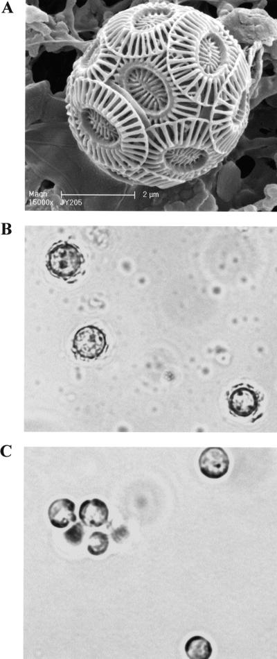
FIG. 1.
(A) Scanning electron micrograph of coccolith-bearing cell of E. huxleyi CCMP1516 showing overlapping coccoliths (magnification, ×15,000; courtesy of J. R. Young). (B and C) Light microscopy images of CCMP 1516 cells showing (B) up-regulation of calcification in phosphate-limited (f/50) medium and (C) down-regulation in phosphate-replete (f/2) medium. Magnification, ×2,000.

