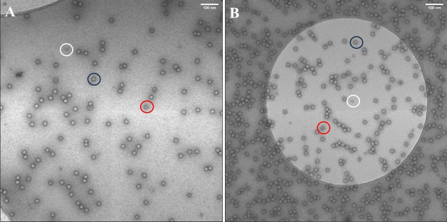
Figure 9. Negative stain transmission electron microscopy images.
A. AAV9-EF1-N-cG produced by suspension 1F11S cells. B. AAV9-EF1-N-cG produced by adherent 1F11 cells. The black circle indicates a full capsid having no darkly stained center. The red circle indicates an empty particle showing a dark stain in the particle center. The white circle indicates impurities previously identified as ferritin (Grieger et al., 2016). Scale bar: 100 nm.

