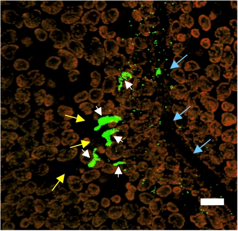
FIG. 4.
Micrograph of GFP-labeled S. enterica serovar Thompson RM1987N cells on the leaf surface of cilantro plants incubated under humid conditions. Large cell aggregates (white arrowheads) are apparent in the vicinity of stomates (yellow arrows) near a leaf vein (blue arrows). Single GFP-S. enterica serovar Thompson cells are also present. The red fluorescent objects are the autofluorescent chloroplasts of the plant epidermal cells. The image is a projected z series obtained with a Leica TCS-NT confocal laser scanning microscope (Leica Microsystems, Wetzlar, Germany). Bar, 20 μm.

