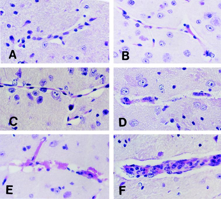
FIG. 3.
Morphology of CM lesions in the brains of C57BL/6J mice. Normal C57BL/6J mice were challenged intravenously with 104 parasitized erythrocytes. Brains were removed for histologic examination at different times. (A) Section of normal mouse brain showing a healthy unaffected blood vessel. (B) Section of mouse brain on day 3 after challenge showing microglial cells in the perivascular space. (C) Section of mouse brain on day 5 after challenge showing increased numbers of activated microglial cells in perivascular spaces. (D and E) Sections of mouse brain on day 7 after challenge showing destruction of endothelial cells and disruption of vessel walls. Endothelial and other cell nuclei were occasionally condensed and/or fragmented in a fashion consistent with apoptosis. (F) Section of mouse brain on day 7 after challenge showing mononuclear cell accumulation and endothelial cell activation in a blood vessel from the brain of a mouse moribund with CM. These were focal lesions. Larger areas of the brains of the same mice demonstrated the changes shown in panels D and E. All sections were stained with H&E. Magnification, ×500.

