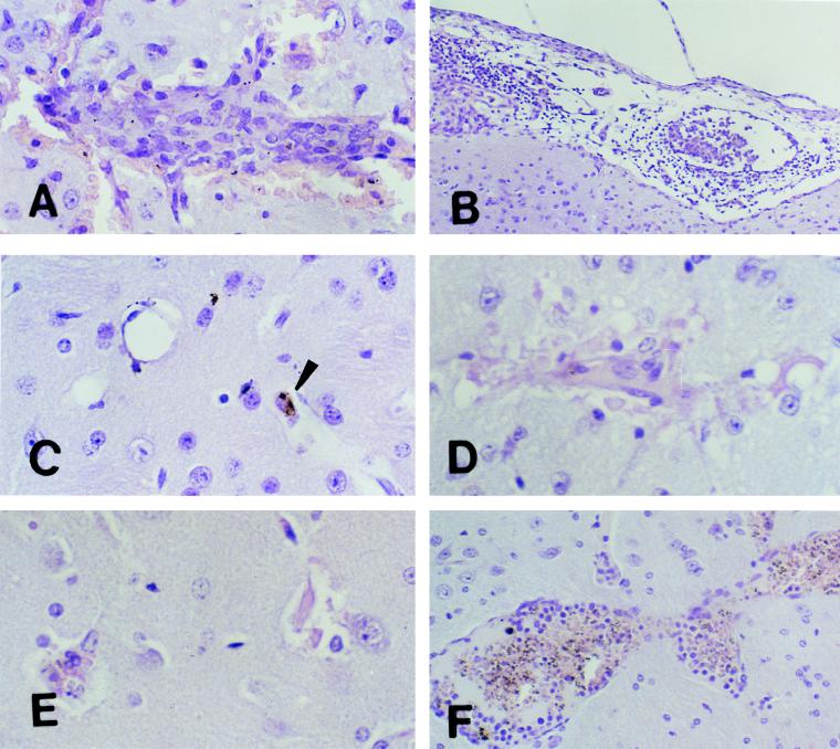
FIG. 5.
Morphologies of lesions in the brains of immunized C57BL/6J (A to C) and A/J (D to F) mice. Immunized C57BL/6J and A/J mice were challenged intravenously with 104 parasitized erythrocytes. C57BL/6J mice were clinically normal, with no signs of CM. In contrast, some A/J mice developed CM while others remained clinically normal, with no signs of CM. Brains were removed from both groups for histologic examination at different times. (A) Section of mouse brain on day 10 after challenge showing an inflammatory hemorrhagic vascular lesion in the cortex. Magnification, ×500. (B) Section of mouse brain and meninges on day 10 after challenge showing meningitis. The monocyte was the predominant inflammatory cell within both vessels and surrounding tissue. Magnification, ×200. (C) Section of mouse brain 21 days after challenge showing resolution and reversibility of the inflammatory and hemorrhagic lesions. Small hemosiderin deposits (arrowhead) are the only remnants of previous lesions. Magnification, ×500. (D and E) Sections of the brains of mice moribund with CM on day 8 after challenge. Changes in microglial cells, destruction of endothelial cells, and disruption of the vessel walls similar to that observed in C57BL/6J mice with CM are evident. The sections appear pale because of loss of cellular integrity. Magnification, ×500. (F) Section of the brain of a mouse protected from lethal CM on day 18 after challenge showing an intense inflammatory infiltrate within blood vessels made up primarily of activated lymphocytes, macrophages, and malarial pigment but in which the endothelium remains intact. These were focal lesions. Areas of the brains of these mice away from these lesions appeared normal. Magnification, ×200. All sections were stained with H&E.

