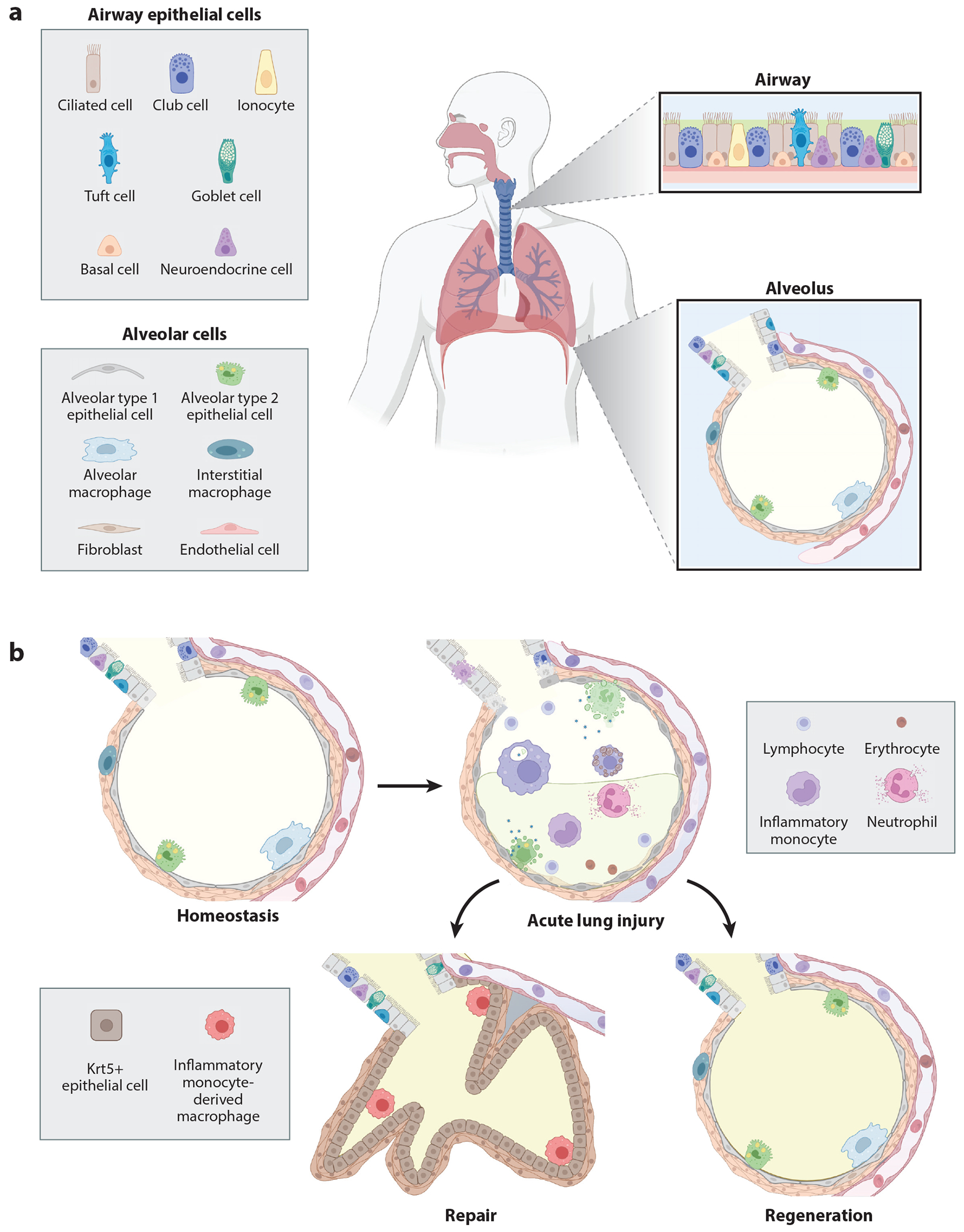
Figure 1.

(a) Epithelium of the lower airway and composition of the alveolus. (b) Repair and regeneration of the alveolus following injury. At homeostasis, the alveolar epithelium consists of squamous alveolar type 1 epithelial cells that are located in close contact with the capillary bed to facilitate gas exchange, as well as cuboidal alveolar type 2 epithelial cells that secrete surfactant stored in lamellar bodies. The alveolus is surrounded by a sparse interstitium composed of fibroblasts, interstitial macrophages, and other cell types not depicted here (e.g., lymphatic vessels and nerves). Viral injury causes alveolar epithelial cell death, barrier dysfunction, impaired gas exchange, alveolar hemorrhage, and infiltration of leukocytes and protein-rich fluid. Resolution of inflammation can occur via regeneration (reconstitution of the functional alveolus) or repair (scarring). Repaired epithelium does not participate in gas exchange and contains airway-derived cuboidal epithelial cells expressing Krt5+. Abbreviation: Krt5, keratin 5. Figure adapted from images created with BioRender.
