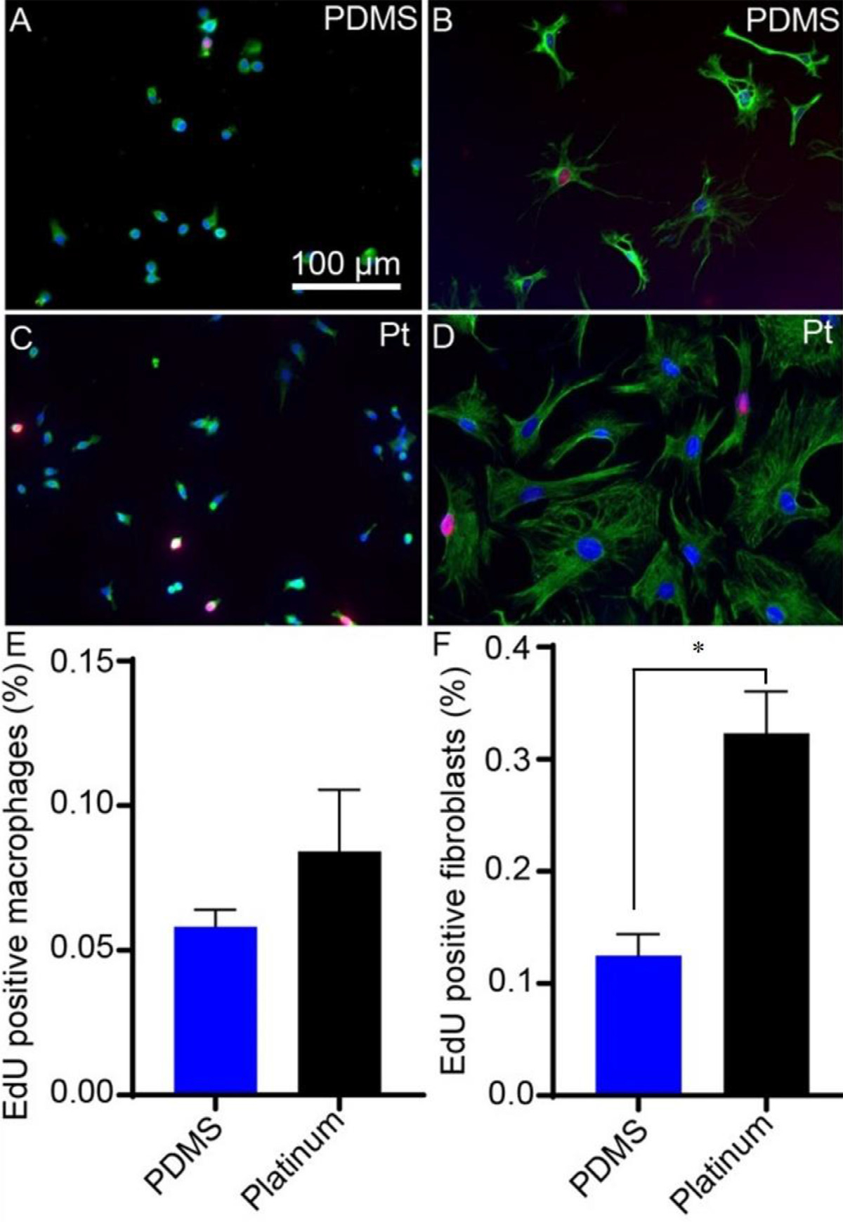
Fig. 3.

Cell proliferation on PDMS and platinum surfaces. Macrophages and fibroblasts were cultured on PDMS and platinum. Macrophages (A,C) were labeled with anti-F4/80 antibody (green) and DAPI (blue). Fibroblasts (B,D) were labeled with anti-vimentin antibody (green) and DAPI (blue). Cell proliferation was measured as the percentage of EdU-expressing (red) nuclei. Macrophage proliferation was similar on PDMS and platinum surfaces (E), p = 0.30. Fibroblast proliferation differed significantly, with a greater percentage of EdU-expressing fibroblasts in the platinum condition relative to PDMS (F), p = 0.030. The experiment was repeated with at least 3 separate cultures. Error bars present standard error of the mean.
