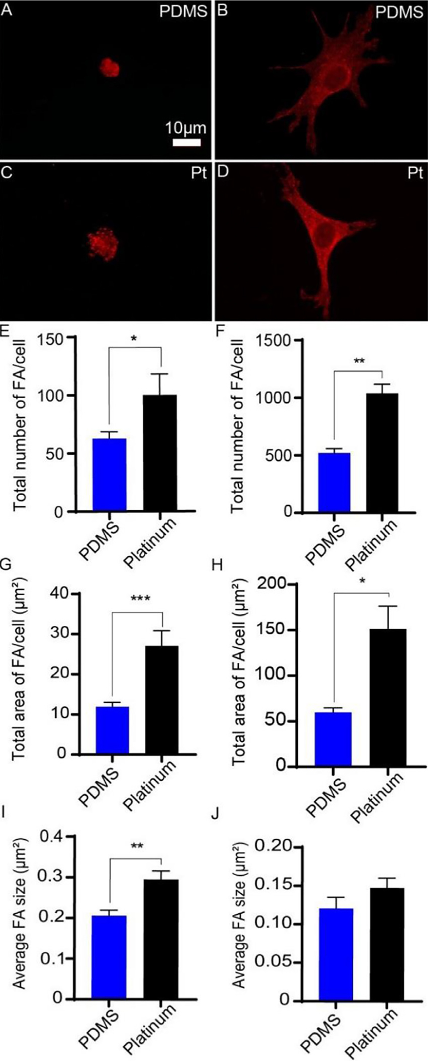
Fig. 4.

Focal adhesion formation on PDMS and platinum surfaces. Macrophages (A,C) and cochlear fibroblasts (B,D) were cultured on PDMS and platinum for 4 h, followed by labeling with anti-phosphorylated FAK antibody (red). Macrophages formed a greater number of focal adhesions on platinum surfaces relative to PDMS (E), p = 0.031, and the focal adhesions were larger on average (I), p = 0.003, and occupied a greater surface area on platinum than on PDMS (G), p = 0.0004. Similarly, fibroblasts formed a greater number of focal adhesions on platinum relative to PDMS (F), p = 0.004. The focal adhesions occupied a greater surface area on platinum (H), p = 0.023, however there were no differences in the mean size of the focal adhesions between fibroblasts grown on PDMS and platinum (J), p = 0.24. The experiment was repeated with at least 3 separate cultures. Error bars present standard error.
