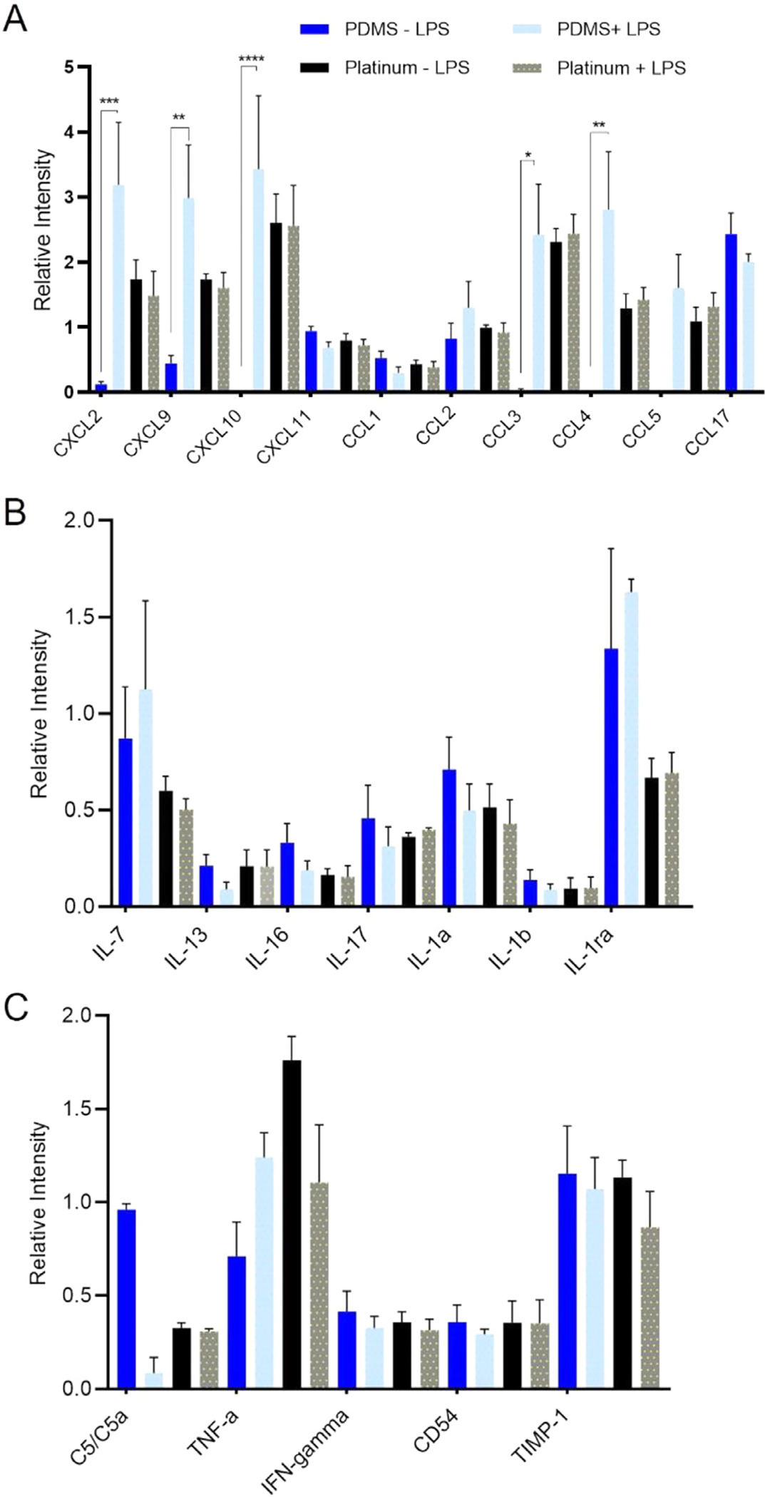
Fig. 5.

Cytokine expression on PDMS and platinum surfaces. Macrophages were cultured on PDMS and platinum for 7 days, then underwent an endotoxin challenge with the addition of 25 ng/mL of lipopolysaccharide (LPS) for 8 hrs to prime the cells. Medium without LPS was used as a control. After 8 h, 1.5 mL of medium from each condition was collected for cytokine analysis. Chemokine/cytokine (A), interleukin (B), and other pro-inflammatory marker (C) expression is represented as a relative intensity. In the PDMS condition, increased levels of CXCL2 (p = 0.0001), CXCL9 (p = 0.007), CXCL10 (p < 0.0001), CCL3 (p = 0.019), and CCL4 (p = 0.001) were expressed when the macrophages were primed with LPS. Expression levels of interleukins and other pro-inflammatory markers were similar between LPS and non-LPS groups. Cytokine/chemokine, interleukin, and other pro-inflammatory marker expression from macrophages grown on platinum did not differ between LPS and non-LPS groups. When comparing PDMS and platinum groups without LPS, CXCL10 (p = 0.005) and CCL3 (p = 0.038) were the only chemokines/cytokines with increased expression in the platinum conditions relative to PDMS. When comparing PDMS and platinum groups with LPS, IL-1ra was the only interleukin with significantly higher expression on PDMS relative to platinum (p = 0.044). The experiment was repeated with at least 3 separate cultures. Error bars present standard error.
