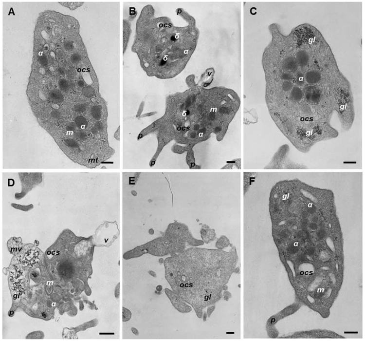
Fig. 3. Representative transmission electron micrographs of human platelets without or with characteristic ultrastructural alterations induced by anti-dsDNA Abs or anti-dsDNA Abs/dsDNA complexes.
Ultrastructure of resting (A, F) and activated (B, C, D, E) platelets: (B) shape change, shrinkage and formation of multiple filopodia; (C) shape change, α-granules grouped in the middle of the platelet body; (D) dramatic shape change, partial fragmentation, a solid electron dense area in the center, vesiculation of the plasma membrane; (E) a “grey” platelet without α-granules. Designations: α, α-granules; δ, δ-granules, gl, glycogen granules; m, mitochondria; mt, microtubules; OCS, open canalicular system; f, filopodia; v, microvesicle with a single membrane; mv, a multivesicular particle. Scale bars are 0.2 μm (A, B, D, E) and 0.5 μm (C).

