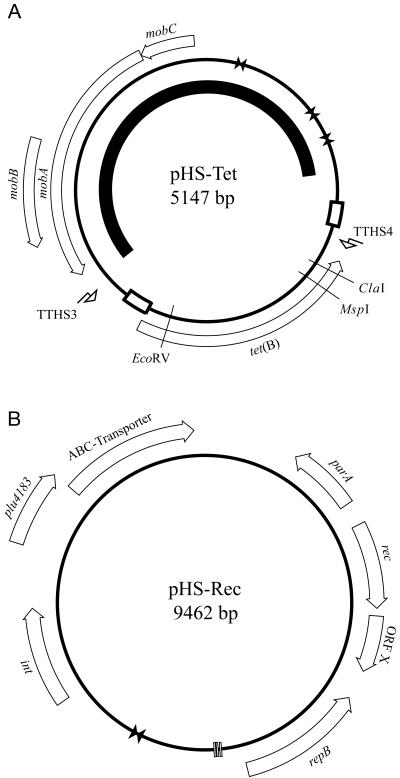
FIG. 1.
Schematic map of plasmid pHS-Tet (A) and pHS-Rec (B). White arrows indicate putative ORFs, vertical bars indicate direct repeats, bow ties (opposing triangles) indicate inverted repeats, and bent arrows indicate primer binding sites. The black arc inside the map of pHS-Tet indicates the region of pHS-Tet sharing similarity with plasmid pAB2. The white boxes on the map of pHS-Tet indicate regions of duplicated sequence.

